
Customizable Whole-Body MRI Cancer Screening
(DWIBS Method and More)
〜Do you have any concerns about your body?〜
Our clinic combines a Whole-Body MRI Cancer Screening that thoroughly checks the entire body with a more precise MRI Breast Cancer Screening, allowing us to offer a more accurate screening tailored to each person’s individual risk. For those who want to carefully examine areas of concern, or who want a screening method that places as little burden on the body as possible, we propose a new form of cancer screening that you can continue with peace of mind.
Menu
Whole-Body MRI Cancer Screening
Our Whole-Body MRI Cancer Screening is based on the imaging theory developed by Dr. Taro Takahara, the originator of the DWIBS (Diffusion-Weighted whole-body Imaging with Background body signal Suppression) method. Building on this foundation and drawing on expertise specialized in cancer care, we provide a fully “custom-tailored” screening in which imaging conditions are optimized for each individual patient’s condition.
Target Diseases
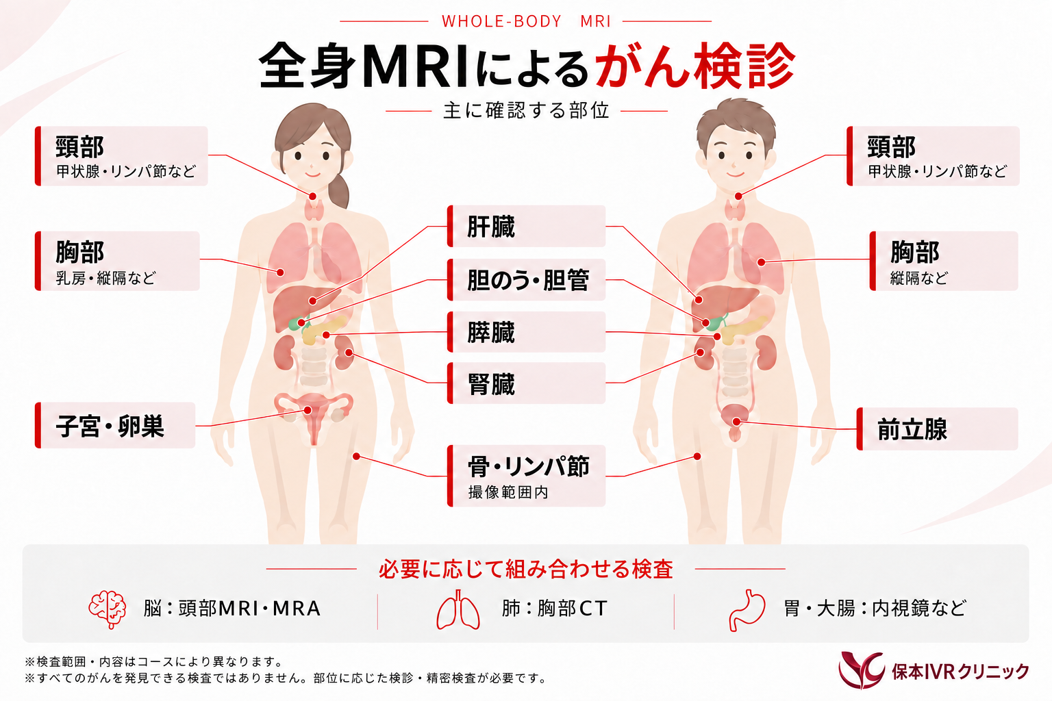
Features of Whole-Body MRI Cancer Screening
- Low burden on the body: No radiation exposure or injections.
- Detects conditions beyond cancer: Identifies a variety of abnormalities, not just cancer, at the same time.
- No restrictions after the exam: You can return to daily activities such as bathing and eating immediately after the exam.
- Suitable for people with diabetes: Since there are no blood-glucose-related restrictions, you can undergo the exam with peace of mind.
- Thorough examination: The exam takes about 45 minutes. We combine multiple imaging methods to carefully check for cancer and other conditions from multiple angles, taking the time needed for a thorough examination.
Why There Is Zero Radiation Exposure
As the name suggests, Whole-Body MRI Cancer Screening uses MRI. An MRI is a device that takes images of the body while you lie inside a tunnel-like scanner.
Because the inside of an MRI machine consists of magnets and coils and does not use X-rays, radiation exposure is “completely zero.”
This means you can undergo the exam repeatedly with peace of mind.
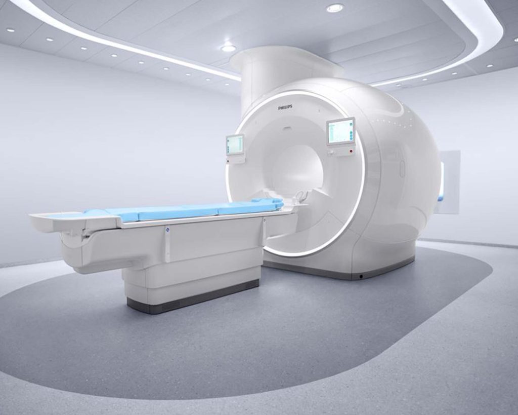
This is a state-of-the-art, helium-free MRI system — Philips’ top-grade medical imaging diagnostic equipment. It is equipped with “DUAL AI (SmartSpeed Precise),” one of the world’s most advanced high-definition imaging technologies, which uses artificial intelligence in two complementary ways to deliver dramatically faster scans compared with conventional systems. By thoroughly removing noise, it achieves both improved image quality and faster imaging at the same time. This is a powerful tool for helping determine treatment strategies and for the early, radiation-free detection of cancer. In addition, our clinic is equipped with a special video system that projects beautiful imagery onto the wall via a mirror during the scan, allowing even those who find the narrow space inside the MRI uncomfortable to undergo the exam while relaxed.
During the MRI exam, rather than a purely clinical setting, you can undergo the scan in a calm environment filled with beautiful imagery and music.
This helps ease patients’ anxiety and tension, providing a more comfortable, higher-quality examination experience.
The DWIBS Method
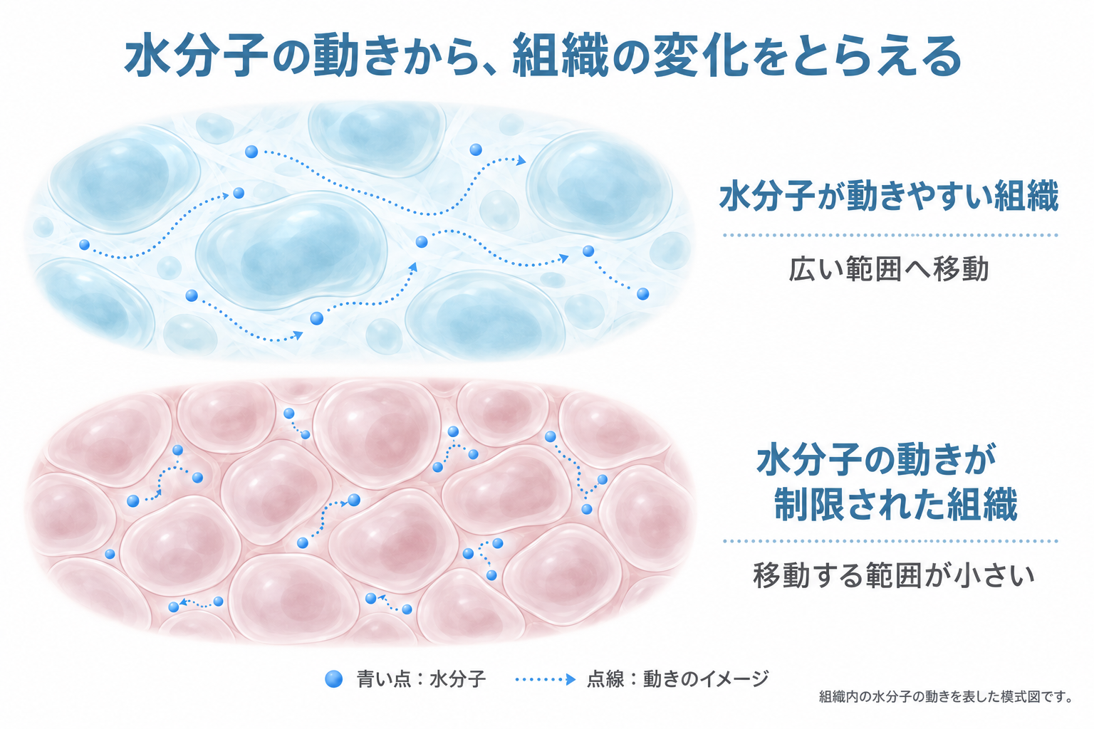
This is a specialized imaging technique that visualizes the movement of water molecules within cells. In normal cells, water molecules move actively, but in areas affected by tumors, inflammation, or infarction, cell density increases and the movement of water molecules becomes restricted. The DWIBS method detects lesions by capturing this difference in molecular movement. The imaging range broadly covers the area from the level of the ears down to the pelvis.
Whole-Spine Imaging
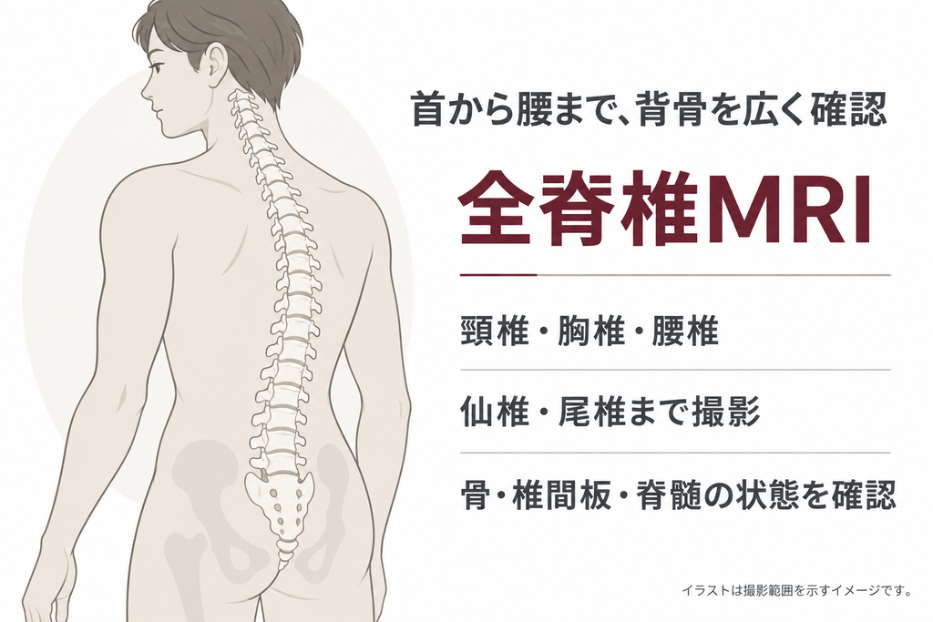
This images the entire spine (cervical, thoracic, lumbar, sacral, and coccygeal vertebrae) to identify spinal conditions such as compression fractures caused by osteoporosis, herniated discs, and spinal stenosis. It is useful for those concerned about spinal misalignment, neck and shoulder stiffness, or lower back pain. It can also detect bone-related conditions such as ligament calcification, bone hyperplasia, tumors, and cancer metastasis.
Abdominal Imaging (MRCP)
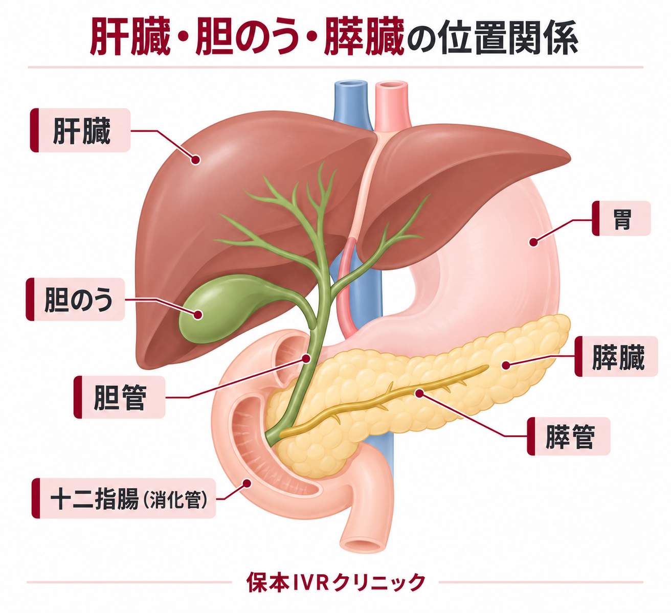
This images the abdominal organs (liver, pancreas, kidneys, gallbladder, spleen, and digestive tract). It also includes a technique called “MRCP,” which allows detailed visualization of the pancreatic duct and bile duct.
Pancreatic cancer and biliary tract cancer (gallbladder cancer and bile duct cancer) are both known to have a poor prognosis, and in most cases they first cause abnormalities in the pancreatic duct or bile duct — by the time symptoms appear, it is often already too late. By capturing these abnormalities, MRCP contributes to the early detection of pancreatic cancer and biliary tract cancer.
In particular, the incidence of pancreatic cancer is increasing in both men and women, and this disease is easily missed by ultrasound or non-contrast (plain) CT.
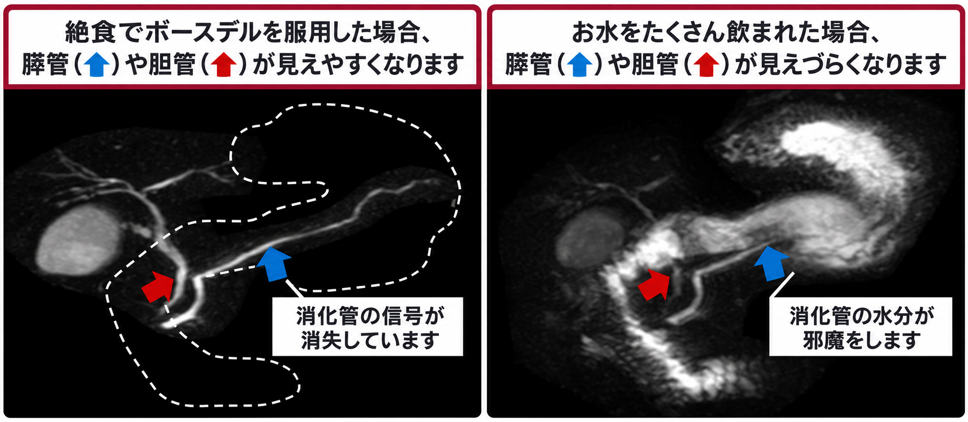
※When imaging the abdominal organs, residual food in the stomach or fluid remaining in the duodenum can interfere with diagnosis. For this reason, you will be asked to fast for 6 hours before the exam. You will also be asked to drink an oral contrast agent (Boursdel) just before the exam. Taking Boursdel allows the pancreatic duct and bile duct to be visualized more clearly.
Pelvic Imaging
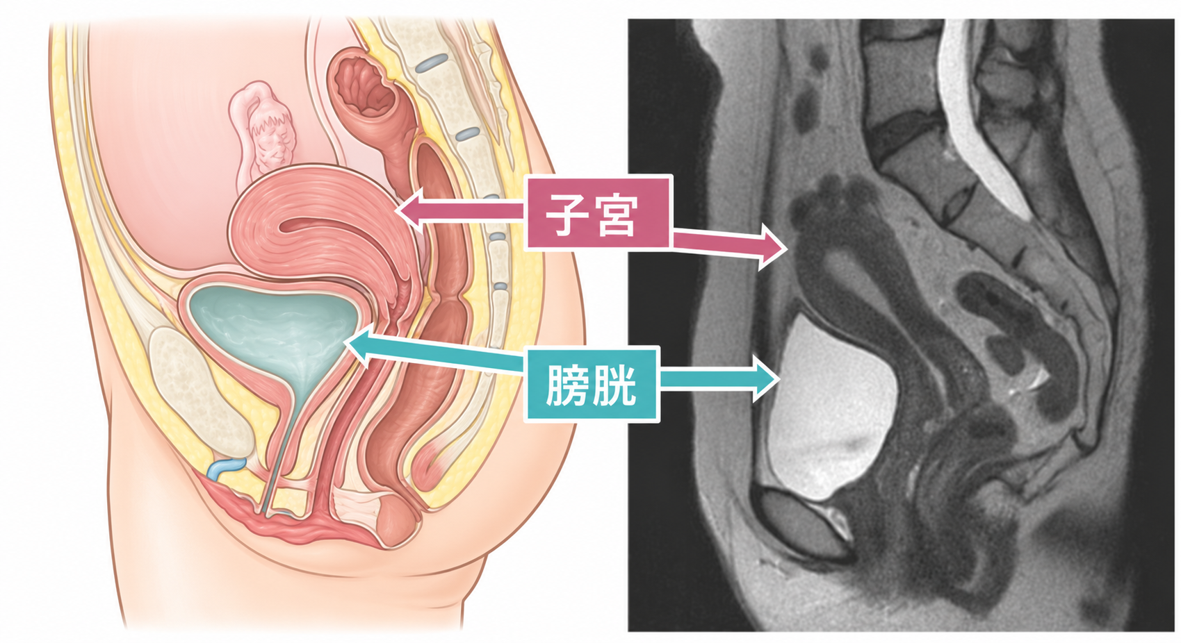
This images the pelvic organs (bladder, lower digestive tract, and — for women — the uterus and ovaries, or for men — the prostate). In addition to conditions of the bladder and lower digestive tract, it can detect uterine and ovarian conditions in women (internal and external uterine abnormalities, cervical cancer, endometrial cancer, uterine fibroids, and ovarian cancer), and in men, conditions such as prostate cancer and benign prostatic hyperplasia.
Screening Flow
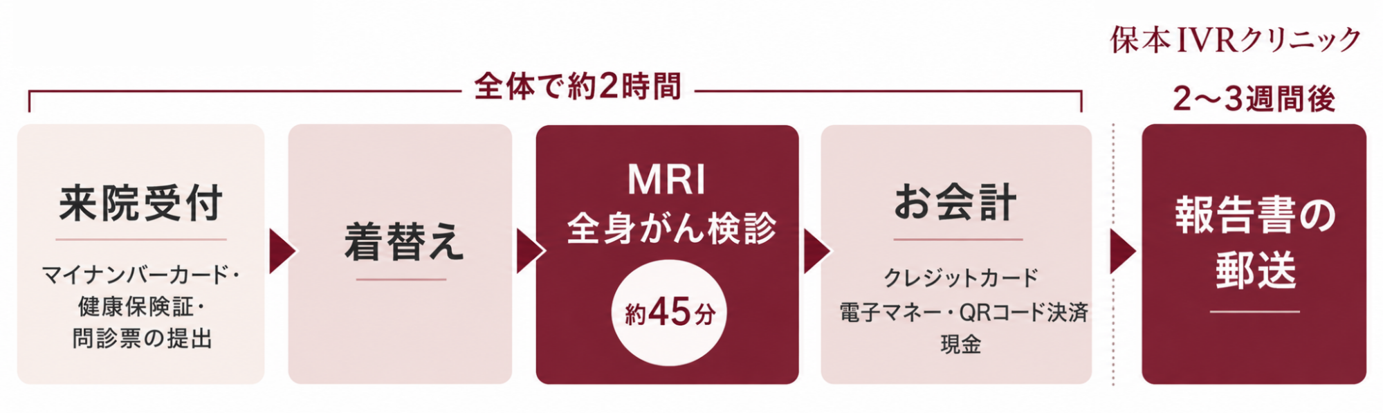
Examination Fees
Whole-Body MRI Cancer Screening
| Screening Content | Excl. Tax | Incl. Tax |
|---|---|---|
| Whole-Body MRI Cancer Screening | ¥95,000 | ¥104,500 |
Fees for Examinations Other Than Whole-Body MRI Cancer Screening
| Screening Content | Excl. Tax | Incl. Tax |
|---|---|---|
| MRI Breast Cancer Screening | ¥23,000 | ¥25,300 |
| Chest CT Scan | ¥26,000 | ¥28,600 |
| Brain MRI Scan | ¥30,000 | ¥33,000 |
| General Blood Test | ¥6,000 | ¥6,600 |
| Tumor Marker (per marker) | ¥2,000 | ¥2,200 |
※If you would like to add any of the above as an option to the Whole-Body MRI Cancer Screening, please contact us.
About the Platinum Screening Plan
We also offer a “Platinum Screening Plan,” which combines “whole-body MRI (including MRCP, brain MRI, and painless breast cancer screening)” with “plain CT or whole-body contrast-enhanced CT” and “blood tests,” and includes an “interpretation and explanation” of the results by Director Yasumoto, based on approximately 5,000 high-resolution images.
For specific inquiries, please contact Best Medical Club Co., Ltd. (BMC), our partner organization.
Reservations
By Phone
Please Call Us First
Reception Hours
- Mon–Fri: Morning 9:00–12:00 / Afternoon 13:00–17:00
- 1st, 3rd, 5th Saturdays: Morning 9:00–12:00
Closed Days
- Sundays, 2nd & 4th Saturdays, national holidays, year-end and New Year holidays
Frequently Asked Questions
Q. What is the difference between Whole-Body MRI Cancer Screening (DWIBS method) and PET-CT?
The imaging principle (examination method) is different. Whole-Body MRI Cancer Screening (the DWIBS method) is a technique that takes advantage of the high cell density of malignant tumors, whereas PET-CT is a technique that takes advantage of the high glucose metabolism of malignant tumors. Because of this difference, there are also differences in lesion detection.
Specifically, Whole-Body MRI Cancer Screening can have difficulty visualizing and detecting lesions around the lungs and heart. PET-CT, on the other hand, tends to accumulate the tracer in urinary tract tissue, so the urinary tract is detected even when normal, which can make it difficult to detect lesions nearby. The table below summarizes other differences.
| Whole-Body MRI Cancer Screening | PET-CT | |
|---|---|---|
| Injection | None | Intravenous injection of a tracer |
| Test Agent | Oral contrast agent | Radiopharmaceutical |
| Dietary Restrictions | Fasting before the exam※ | Fasting and insulin restrictions before the exam |
| X-ray–Related Damage | None | Radiation exposure occurs |
| Exam Duration | Approx. 40 minutes | Approx. 3 hours including the injection |
| Post-Exam Care | None | Must wait until radioactivity decreases |
| Diabetic Patients | Can be examined | May not be able to be examined |
※ Because fasting is required for Whole-Body MRI Cancer Screening, patients with diabetes are restricted from administering insulin injections before the exam.
Q. What conditions are difficult to detect with Whole-Body MRI Cancer Screening?
Lesions near the lungs and heart can be difficult to detect. If you wish to examine the chest in detail, we recommend adding a chest CT scan as an option. Also, since Whole-Body MRI Cancer Screening covers the imaging range from the level of the ears to the pelvis, the head (brain) and limbs are outside the imaging range. Areas not included in the imaging range can be examined with additional optional tests, so please see here for details.
Q. I have claustrophobia — can I still undergo the exam?
To help patients stay relaxed during the exam, you can listen to music while the scan is underway. In addition, our technologist monitors your condition and will check in with you as needed. You will be given a buzzer to hold, so please press it immediately if you begin to feel uncomfortable during the exam. If you do, we will stop the exam and the technologist will come into the exam room to assist you.
Q. Is the breast included in the imaging range of Whole-Body MRI Cancer Screening?
Yes, the breast is included. However, the detection capability for breast lesions with Whole-Body MRI Cancer Screening is about 1 cm, so for those who want a more detailed breast examination, we recommend the MRI Breast Cancer Screening. Because the MRI Breast Cancer Screening uses equipment dedicated to breast imaging, it can detect breast lesions as small as about 5 mm. If you would like to have both a whole-body and a breast examination, we recommend adding the breast cancer screening as an option — please see here for details.
Q. What conditions are difficult to detect with Whole-Body MRI Cancer Screening?
Lesions near the lungs and heart can be difficult to detect. If you wish to examine the chest in detail, we recommend adding a chest CT scan as an option. Also, since Whole-Body MRI Cancer Screening covers the imaging range from the level of the ears to the pelvis, the head (brain) and limbs are outside the imaging range. Areas not included in the imaging range can be examined with additional optional tests, so please seehere for details.
Q. What should I be careful about when taking the oral contrast agent (Boursdel)?
Because the oral contrast agent (Boursdel) contains manganese, it may rarely cause loose stools or diarrhea. In addition, the following individuals may be unable to take Boursdel, or may only be able to take it under certain conditions. Please be sure to inform our staff before the exam.
- Boursdel cannot be used for the exam
- Have previously experienced an allergic reaction after taking Boursdel.
- Have had gastrointestinal perforation, or have undergone surgery on the stomach, duodenum, gallbladder, pancreatic duct, etc., and have been told there is a suspected gastrointestinal perforation.
- Boursdel can be used for the exam under certain conditions
- Have been told by your primary care physician to restrict fluid intake, or are undergoing dialysis. (Boursdel contains 250 mL of liquid, so please be aware that you will need to consume this amount of liquid as part of the Whole-Body MRI Cancer Screening.)
- Are taking oral tetracycline antibiotics, oral new quinolone antibacterial agents, or cefdinir. (Because these medications’ effects may be weakened, you will need to wait about 3 hours after taking the contrast agent before taking these medications.)
If you have any questions about the above or anything else, please feel free to contact us.
Q. What kind of sound does the MRI make?
Due to the nature of MRI, it makes a loud noise during scanning. Once you get used to it, it is not much of an issue, but some people may find it unpleasant the first time. We have uploaded a sample of the sound to YouTube, so if you are concerned, please listen to it beforehand.
Q. Are there cases where I cannot undergo an MRI exam?
There are cases where patients cannot undergo an MRI exam due to medical devices inside or on the body, or due to being in poor physical condition. In addition, it is generally considered best for pregnant women (up to 14 weeks of pregnancy) to avoid the exam.
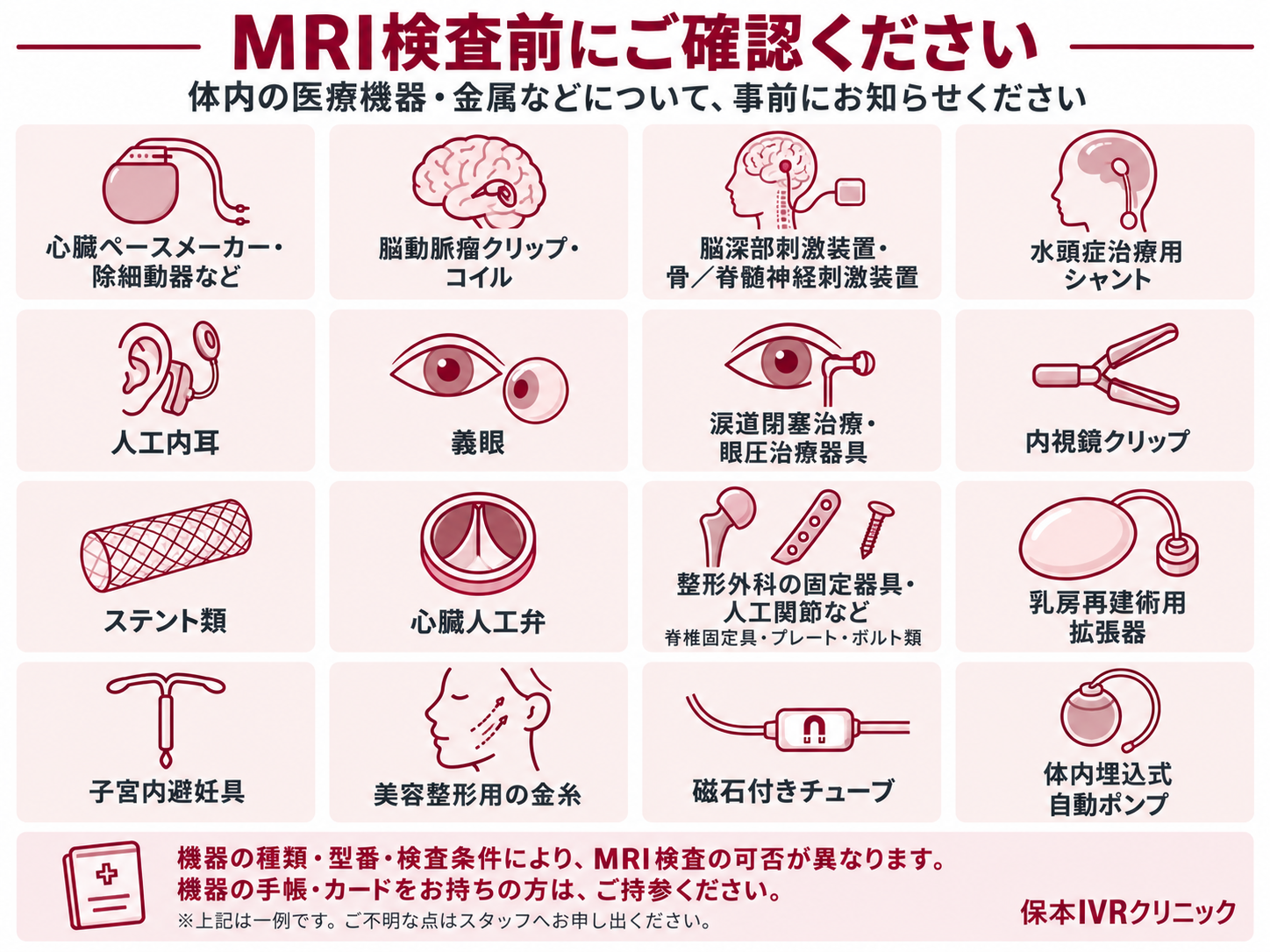
Please see below for examples of medical devices and items that must be removed before an MRI exam. If you have any questions, please feel free to contact us.
Please inform us in advance if you have any of the medical devices listed below, or any other medical devices
- Cardiac pacemakers, defibrillators, etc.
- Cerebral aneurysm clips, coils
- Deep brain stimulators, bone/spinal cord nerve stimulators
- Hydrocephalus shunts
- Cochlear implants
- Artificial eyes
- Lacrimal duct obstruction treatment devices, intraocular pressure treatment devices
- Endoscopic clips
- Stents
- Artificial heart valves
- Orthopedic devices (spinal fixation devices, artificial joints, plates, bolts, etc.)
- Breast reconstruction tissue expanders
- Intrauterine devices (IUDs)
- Cosmetic gold thread implants
- Tubes with magnetic caps
- Implanted automatic pumps
You will be asked to remove the following personal items
- Hearing aids
- Dentures
- Contact lenses
- Disposable body warmers
- Medicated patches/plasters
- Glasses
- Acupuncture needles
- Wigs/hairpieces
- Nail art/artificial nails
- Accessories/jewelry
- Hairpins
- Watches
- Electronic devices and magnetic cards
- Keys
- Metal zippers
- Underwire/metal hooks on undergarments
- Wallets

If you have any questions about the above or anything else, please feel free to contact us.


