

Painless MRI Breast Cancer Screening
~ The new breast cancer screening that doesn’t hurt ~
What Is MRI Breast Cancer Screening?
An entirely new type of breast cancer screening performed with MRI — no contrast agent and no radiation exposure.

Benefits of MRI Breast Cancer Screening
- Unlike conventional mammography, the breast is not compressed, so there is no “pain from compression.”
- You can be examined while wearing a thin layer of clothing (such as a T-shirt).
- There is no radiation exposure.
- The examination takes about 20 minutes.
Features of Painless MRI Breast Cancer Screening
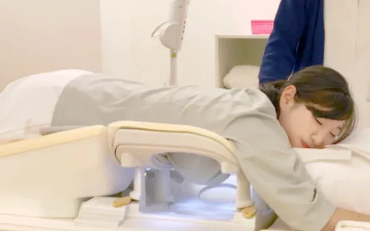
This is an entirely new type of breast cancer screening performed with MRI, without any contrast agent and without any radiation exposure.
- Unlike conventional mammography, the breast is not compressed, so it “doesn’t hurt.”
- You can be examined while wearing a thin layer of clothing (such as a T-shirt).
- There is no radiation exposure.
- The examination takes about 20 minutes.
The Difference Between Painless MRI Breast Cancer Screening and Mammography
- It can visualize areas that are blind spots for mammography, such as the back of the breast (chest wall) and the armpit.
- More than half of Japanese women have abundant breast tissue (dense breasts). This screening can be performed without being affected by breast density.
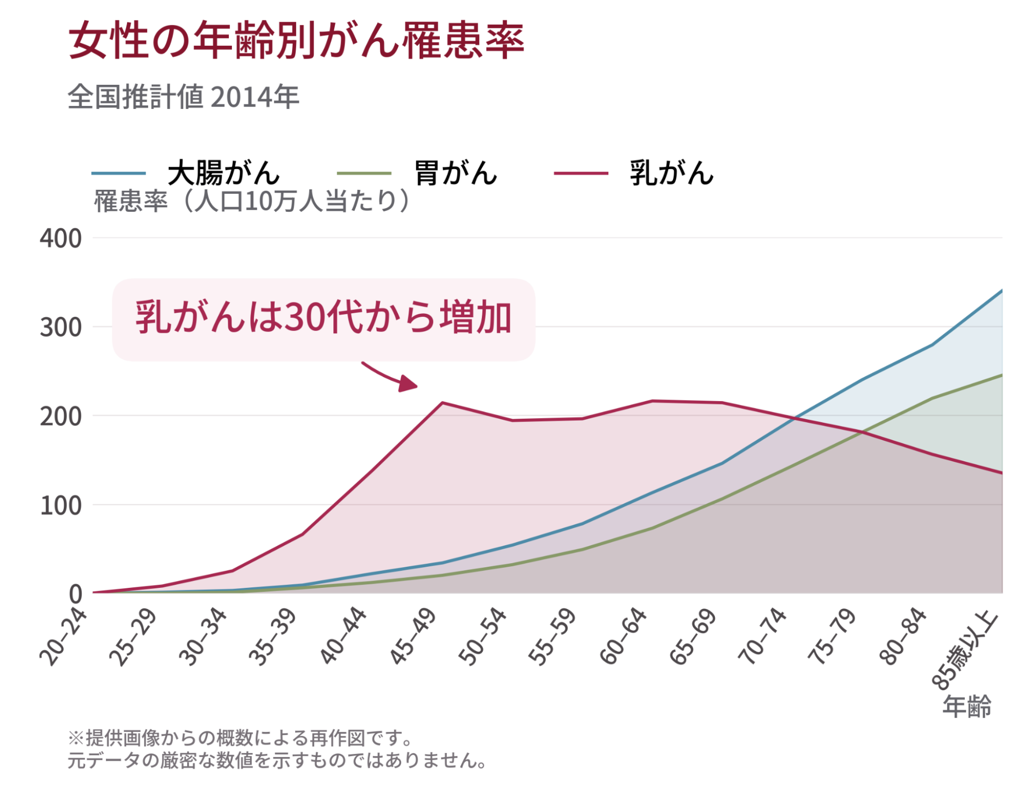
- Mammography is not recommended for women under 40 (to avoid radiation exposure), but this screening method can also protect women in their 30s, when the incidence of breast cancer begins to rise.
Why It Doesn’t Hurt
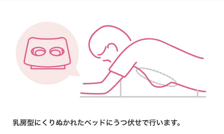
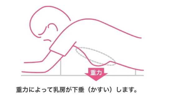
Painless MRI breast cancer screening (DWIBS-Search) is performed lying face down, with the breasts hanging naturally into a round opening to be imaged. Because the breast keeps its natural shape and is never compressed, there is no pain at all — none of the pain associated with having the breast squeezed.
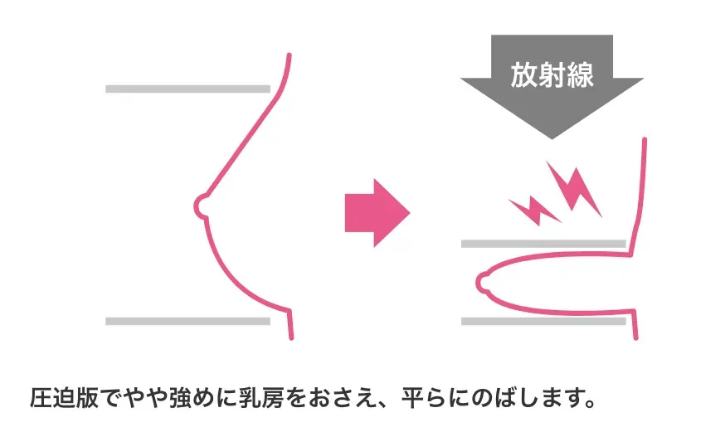
With mammography
With DWIBS-Search
Why There’s No Need to Undress
Mammography requires you to undress for the examination, but with painless MRI breast cancer screening (DWIBS-Search) you can keep your examination gown or T-shirt on. Because the scan uses magnetism to image the body’s organs and blood vessels, wearing a T-shirt does not affect the resulting images.
In addition, the only time the technologist and the patient are in close proximity is when entering and leaving the room. Your privacy is fully protected, so please relax during the examination.
With mammography

With DWIBS-Search
Why There’s Zero Radiation Exposure
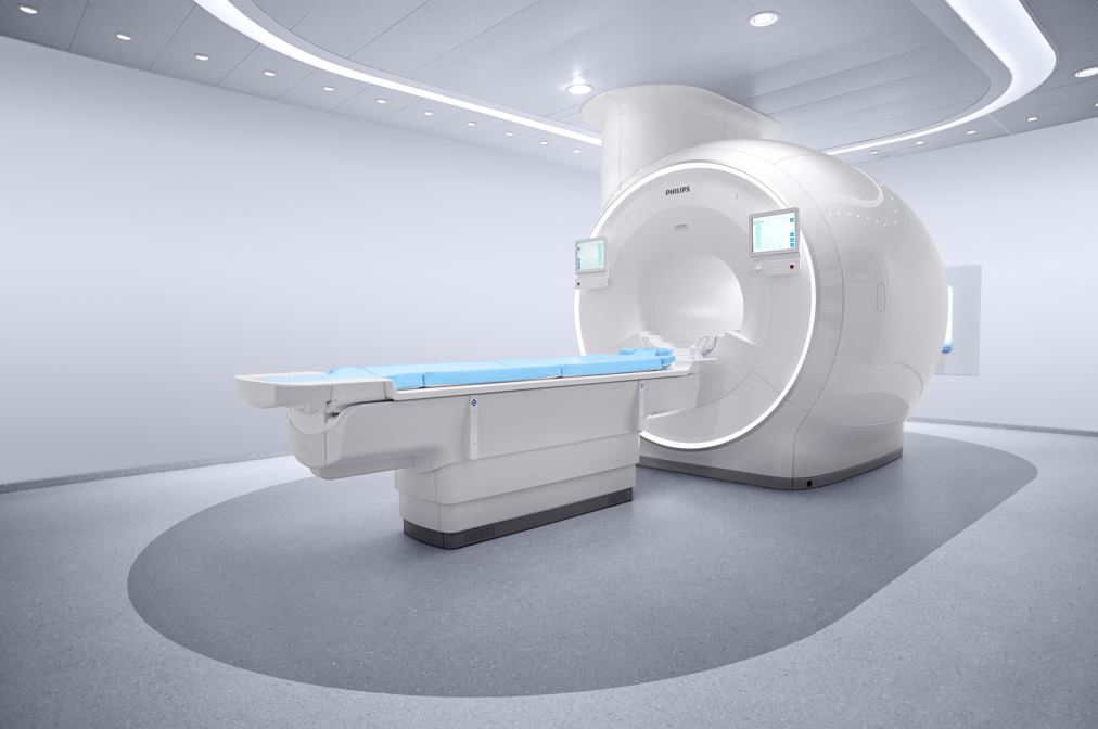
As its name suggests, painless MRI breast cancer screening (DWIBS-Search) uses MRI. MRI is a device that takes images of the body by placing it inside a tunnel-like structure.
The inside of an MRI machine consists of magnets and coils, and because it does not use X-rays, radiation exposure is “completely zero.”
This means you can undergo the examination repeatedly with complete peace of mind.
Why the Cancer Detection Rate Is High
One of the greatest strengths of painless MRI breast cancer screening (DWIBS-Search) is how easily it detects cancer.
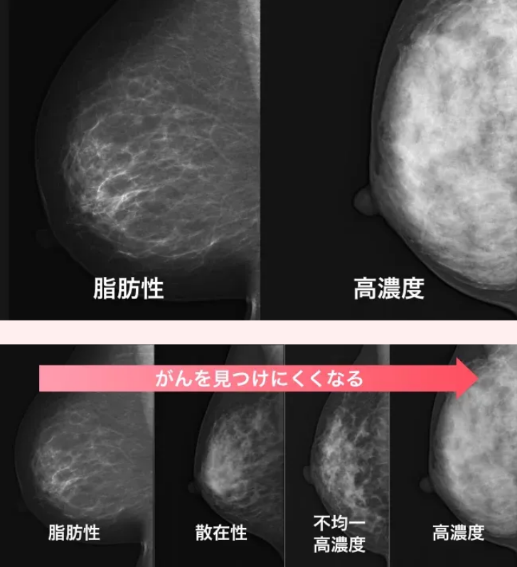
Most women up to around age 50 have abundant breast tissue, which is difficult for mammography’s X-rays to penetrate, causing the center of the breast to appear white (high density) on the image. This is called a “dense breast.”
With mammography, cancer also appears white, so finding it is like trying to spot a “white rabbit in the snow” — naturally making cancer harder to detect.
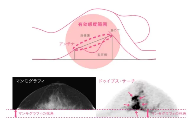
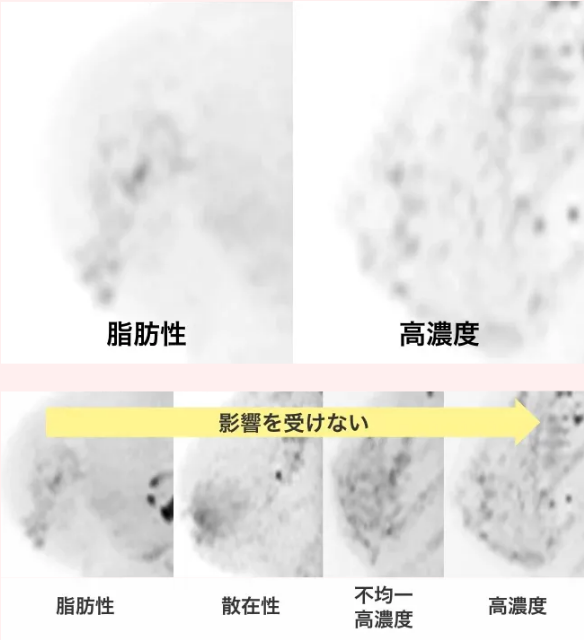
With painless MRI breast cancer screening (DWIBS-Search), cancer appears black, so cancers that are difficult to distinguish on mammography are being found one after another.
In addition, painless MRI breast cancer screening (DWIBS-Search) images the breast evenly, all the way to the back of the breast (chest wall) and the armpit.
MRI has a wide effective sensitivity range, allowing the examination to cover the back of the breast (chest-wall side) and the armpit as well.
Why You Can Still Be Screened After Breast Surgery
Breast surgery can be broadly divided into breast cancer surgery and breast augmentation surgery.
In breast cancer surgery, one breast is usually removed, and an implant is then placed to restore its shape.
In breast augmentation surgery, implants are often placed on both sides.
If an implant is compressed strongly, it can rupture and cause inflammation, deforming the breast that was carefully restored.
For this reason, mammography, which compresses the breast, generally cannot be performed.
Ultrasound examination does not forcefully compress the breast, so it can be performed, but because the area behind the implant is difficult to see, some patients are unable to be examined this way.
Painless MRI breast cancer screening (DWIBS-Search) can be performed even on patients who have had implants placed.
Depending on the type of implant, some artifacts (interfering signals) may appear, but in most cases the examination can be performed without any problem.
Why It Is Well Suited to Japanese Women
Compared with Western women, Japanese women have a higher proportion of dense breast tissue, and reports indicate that nearly 80% of women aged 50 and under have dense breasts (dense or heterogeneously dense).
It is known that the cancer detection rate differs by as much as double between breasts that are translucent on mammography (typically in older women) and breasts that appear cloudy white (dense breasts, common among many working-age women).
Because painless MRI breast cancer screening (DWIBS-Search) is less affected by the amount of breast tissue, the detection rate does not decline. Even women with the dense breasts common among Japanese women can undergo the screening with peace of mind.
With mammography
With DWIBS-Search
Mammography was first adopted as a standard breast cancer screening method in Western countries. Because Western data showed it reduced mortality, it was also introduced in Japan.
However, Asian women’s breasts tend to be firmer and smaller than those of Western women. Mammography images the breast by stretching and compressing it flat, and for the type of breast common among Asian women, this stretching can cause significant pain.
With painless MRI breast cancer screening (DWIBS-Search), you simply fit your breast into a breast-shaped opening without any compression, so it is painless and comfortable. In addition, regardless of size, gravity naturally creates a shape that is easy to image.
Examination Flow
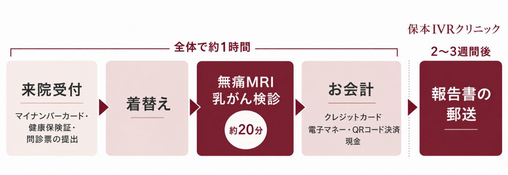
1 Reception
Please check in at our 2nd-floor reception desk, where we will review your medical questionnaire.
2 Change into an examination gown
You will change into an examination gown in the 2nd-floor changing room.
3 Move to the 1st-floor waiting area
We will guide you to the 1st-floor intermediate waiting area.
4 Pre-examination explanation
You will receive an explanation of the examination in the MRI anteroom, and we will review your medical questionnaire.
5 Examination begins
Keeping your examination gown on, you will lie face down with your breast placed in the opening. There is no pain at all.
6 You can press a button if you feel anxious
If you feel anxious partway through, pressing the button will stop the scan immediately.
7 Examination complete
The examination is now complete. Thank you for your patience.
8 Return to the 2nd-floor reception
After changing back into your clothes, please fill out a short questionnaire. Feel free to help yourself to a drink from the 2nd-floor vending machine; a water server is also available.
9 Payment at the 2nd-floor reception
Payment is handled at the clinic’s front reception desk.
If a Suspected Finding Is Detected
We will provide you with the examination results, and you will then undergo a detailed examination at a specialist hospital. Our clinic works closely with many breast cancer specialists and can refer you promptly.
Pricing
Examination Fee
| Screening | Tax included |
|---|---|
| MRI Breast Cancer Screening | ¥25,000 |
Fees for Examinations Other Than MRI Breast Cancer Screening
| Examination | Before tax | Tax included |
|---|---|---|
| Chest CT scan | ¥26,000 | ¥28,600 |
| Brain MRI scan | ¥30,000 | ¥33,000 |
| General blood test | ¥6,000 | ¥6,600 |
| Tumor marker (per marker) | ¥2,000 | ¥2,200 |
Want to Learn More About MRI Breast Cancer Screening?
Please see the link below for more details on MRI breast cancer screening.
MRI Breast Cancer Screening (DWIBS-Search)
Every hospital offering painless MRI breast cancer screening (DWIBS-Search) has its image quality directly adjusted and verified by the physician who developed the “DWIBS method.” Our clinic has also passed these rigorous standards.
Platinum Screening Plan (Platinum Plan)
We also offer a “Platinum Screening Plan,” which combines “whole-body MRI (including MRCP, brain MRI, and painless breast cancer screening)” with either “plain CT or whole-body contrast-enhanced CT” and “blood tests,” providing roughly 5,000 high-resolution images that are reviewed and explained directly by Director Yasumoto.
For specific inquiries, please contact Best Medical Club Co., Ltd. (BMC), a partner of Yasumoto IVR Clinic.
How to Book
Online Booking
MRI Breast Cancer Screening Booking Form
Please book your appointment here
* A cancellation fee may apply depending on circumstances. Please consider carefully before applying for a screening.
We appreciate your understanding and cooperation. Please also note that in some cases you may not be able to undergo the MRI examination.
By Phone
You can also book by phone.
Reception Hours
- Mon–Fri AM/9:00–12:00 PM/13:00–17:00
- 1st, 3rd & 5th Saturdays AM/9:00–12:00
Closed
- Sundays, 2nd & 4th Saturdays, national holidays, year-end/New Year holidays
Frequently Asked Questions
Q. How much time does it take, and do I need to prepare anything?
The examination itself takes about 20 minutes. Including check-in, changing into an examination gown (or simply wearing a T-shirt), and payment, the whole visit takes just over an hour.
No advance rest or injections are required, and there is nothing in particular you need to prepare. Since you can be examined in your gown, there is no dress code. You are welcome to come in work attire, casual outing clothes, or sportswear.
There are no dietary restrictions, and you do not need to stop taking any medications you normally take. You can come exactly as usual. However, you must not wear any magnetic materials on your body.
Q. How much cancer does this screening actually find?
The average cancer detection rate for mammography is 2.7 per 1,000 people (according to 2018 data presented by the Japanese Association of Breast Cancer Screening, based on 2.53 million examinees).
- Tomosynthesis (3D mammography) is said to detect about 1.5 times more cancers.
- Adding ultrasound examination performed by a well-trained technologist to mammography is said to detect about 1.5 times more cancers.
- The cancer detection rate for painless MRI breast cancer screening (DWIBS-Search) was 14.7 per 1,000 people. While a direct comparison is not possible, this corresponds to more than 5 times the mammography rate above.
- The positive predictive value, one of the indicators of a test’s accuracy, was also about 5 times higher.
Q. Does menstruation, pregnancy, or breastfeeding affect the screening?
During the period when the breasts feel swollen before menstruation, the water content of the breast tissue increases, which lowers the ability to detect cancer.
For painless MRI breast cancer screening, we recommend avoiding the first and second days of your period, with days 3–21 recommended (ideally days 5–14 from the start of your period). Even in the 4th week (just before your next period), the examination can still be performed, though not ideally. However, since this is a valuable opportunity for screening, if you want to maximize diagnostic accuracy, we recommend scheduling your visit within the period above.
If your cycle is irregular, please contact the hospital where you made your reservation by phone. Women who have gone through menopause can be examined at any time.
[Regarding breastfeeding and pregnancy]
During breastfeeding, the breast tissue becomes more abundant, which lowers the efficiency of cancer detection. This is why we ask about pregnancy and breastfeeding status in the medical questionnaire. If you choose to be examined under these conditions, please be aware that diagnostic accuracy will be lower than usual.
Please note that MRI during pregnancy requires a physician’s examination, so without a doctor’s recommendation, this screening cannot be performed. (That said, we have received earnest requests from obstetricians and gynecologists: “Some patients notice a lump in the breast after becoming pregnant and are left anxious throughout the pregnancy without any way to find a solution.” We would like to be able to respond to these kinds of concerns.)
Q. Is there any radiation exposure?
None at all. Because this is an MRI examination, radiation exposure is zero. That means your cumulative radiation exposure※ remains at zero no matter how many times you are examined. This makes it an especially suitable test for a health screening you may repeat throughout your life.
It is particularly well suited for people with certain genetic mutations (abnormalities in what are known as the BRCA genes). People with an abnormality in the BRCA gene※※ cannot properly repair DNA damage caused by radiation exposure, which leads to a high rate of cancer development. For this reason, these individuals should avoid X-ray-based examinations (such as mammography or plain chest X-rays) as much as possible.
※ Cumulative radiation exposure (Radiation Cumulative Dose) — the total amount of radiation exposure a person has received over their lifetime.
※※ Hereditary Breast and Ovarian Cancer syndrome (HBOC).
Q. How does MRI breast cancer screening detect cancer?
Cancer tissue is characterized by cells packed tightly together. Because of this, the movement of water (diffusion) within cancer tissue is reduced. Painless MRI breast cancer screening uses this reduced diffusion as a marker to detect cancer. This imaging technique is called “diffusion-weighted imaging.” Among the various approaches, this screening uses a method called DWIBS (Diffusion-Weighted whole-body Imaging with Background body signal Suppression). The resulting images are often said to closely resemble PET scans (FDG-PET), which detect cancer by using the fact that cancer cells consume a lot of glucose. However, since naturally occurring glucose cannot be visualized directly, a radioactive isotope attached to glucose is injected, and its accumulation is then imaged.
Note that this examination (DWIBS-Search) does not use any contrast agent. The gadolinium (Gd)-based contrast agents used in MRI are mostly excreted through urine, but it is known that trace amounts can accumulate in the brain and kidneys. It has also been reported that excreted Gd, a heavy metal, pollutes the environment, with its concentration continuing to rise.
This examination is also well suited for people with a genetic mutation (a BRCA gene mutation, which increases the risk of breast and ovarian cancer) who wish to be screened repeatedly. With contrast-based examinations, concerns about accumulation and cost generally limit screening to about once a year at most; but since this examination uses no contrast agent, is more affordable, and carries no risk of harm or environmental pollution, there is no problem undergoing it, for example, every six months or even three times a year. Shortening the interval between examinations is expected to make it possible to detect cancer within a shorter time after it develops.
Q. How often should I undergo MRI breast cancer screening?
In principle, you should be screened once every two years, ideally once a year. Those considered to be at higher risk of developing breast cancer — for example, women whose mother or sister has had breast cancer — should be screened once a year.
National screening guidelines do not recommend mammography for women under 40, in order to avoid the effects of radiation exposure as much as possible. However, because this examination has nothing to do with radiation, you can undergo it repeatedly with complete peace of mind.
Q. I’m not good with enclosed spaces — will I be okay?
Some people feel tense, sweat, or experience a racing heart when entering a narrow space. However, many people feel more at ease once we explain during the examination that “the tunnel is shorter than your height (about 120 cm).”
Our clinic has introduced Ambient Movie, so that even during the MRI examination, you are not simply in a clinical space but in a calm environment surrounded by beautiful visuals and music. This helps ease patients’ anxiety and tension, providing a more comfortable and higher-quality examination experience.
In addition, many patients feel less anxious because they lie face down for the exam. If it is truly impossible for you, the examination cannot be performed.
Q. Are there cases where I cannot undergo MRI breast cancer screening?
Yes. Because MRI uses a powerful magnet, the following individuals cannot be examined.
- People with large tattoos (some tattoo ink contains iron particles, which can cause burns)
- People wearing a hearing aid (possible if it can be removed)
- People with metal implanted in the chest (may be possible in some cases — please consult with us)
- People with an implanted cardiac pacemaker
In addition, please avoid the following on the day of your examination.
- Spray-type (instant spray) gray hair coloring products (the dispersed iron particles reduce the precision of the MRI equipment; regular hair dye is fine)
- Colored contact lenses or heavy mascara (some contain iron)
Q. What kind of sound does the MRI make?
Due to the nature of the equipment, MRI makes loud noises during scanning. Once you get used to it, it is not much of an issue, but some first-time patients find it uncomfortable. We have uploaded a sample of the sound to YouTube, so if you are concerned, please listen to it in advance.
Q. Are there cases where I cannot undergo an MRI examination?
You may be unable to undergo an MRI examination if you have certain medical devices inside or outside your body, or if you are not feeling well. In addition, it is considered advisable for pregnant women (up to 14 weeks of pregnancy) to avoid the examination.
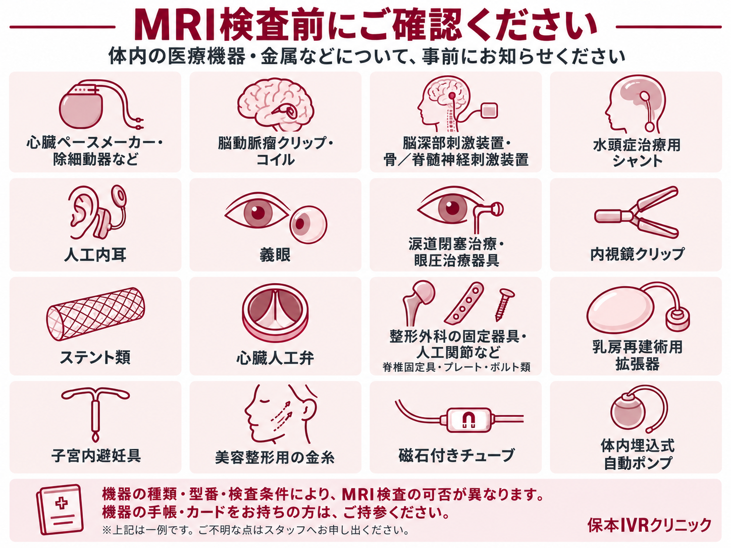
Please see below for examples of medical devices and items that must be removed before an MRI examination. If you have any questions, please feel free to contact us.
Please let us know in advance if you have any of the following medical devices, or any other medical device not listed here
- Cardiac pacemaker, defibrillator, etc.
- Cerebral aneurysm clip or coil
- Deep brain stimulator, spinal cord stimulator
- Shunt for hydrocephalus treatment
- Cochlear implant
- Artificial eye
- Lacrimal duct obstruction device, intraocular pressure treatment device
- Endoscopic clips
- Stents
- Artificial heart valve
- Orthopedic devices (spinal fixation devices, artificial joints, plates, bolts)
- Tissue expander for breast reconstruction
- Intrauterine device (IUD)
- Gold thread used in cosmetic procedures
- Tubes with magnetic caps
- Implanted automatic infusion pump
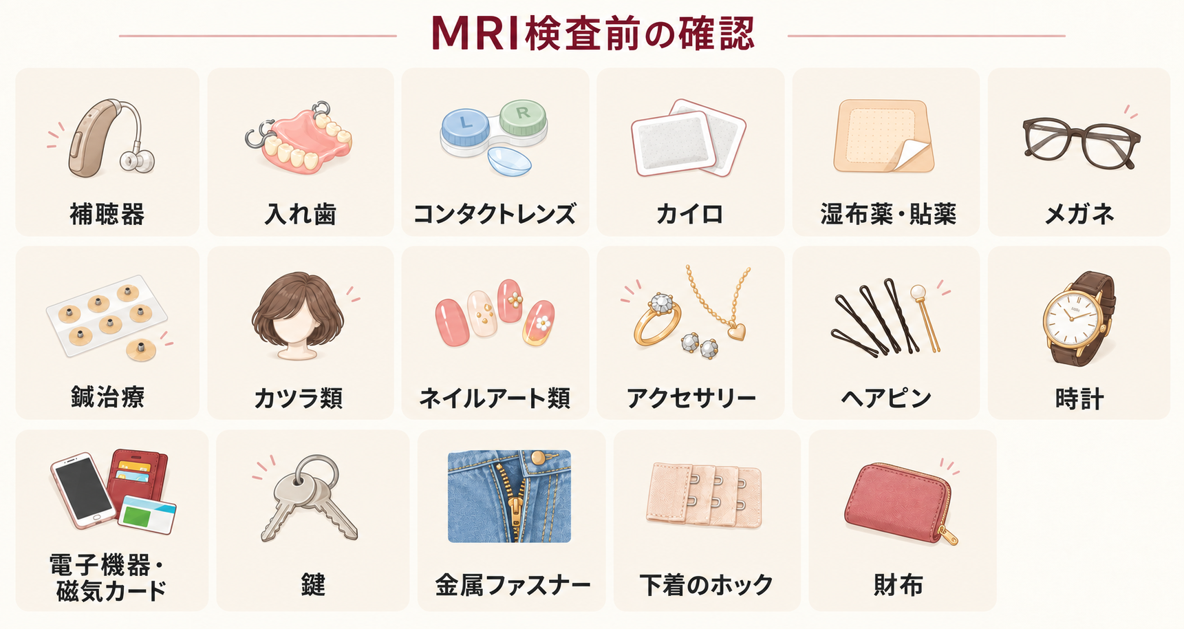
Please remove the following belongings before the examination
- Hearing aid
- Dentures
- Contact lenses
- Disposable body warmers
- Medicated patches/plasters
- Glasses
- Acupuncture needles
- Wigs
- Nail art
- Accessories
- Hairpins
- Watch
- Electronic devices / magnetic cards
- Keys
- Metal zippers
- Underwire/metal hooks on undergarments
- Wallet
If you have any questions about the above or anything else, please feel free to contact us.


