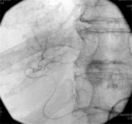
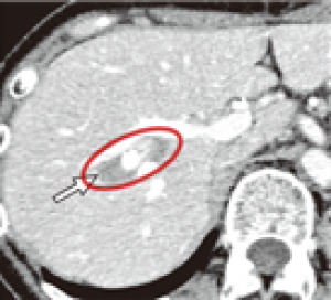
When malignant, liver tumors are broadly classified into "primary liver tumors" (primary liver cancer and bile duct cancer), which arise directly from liver tissue or the bile ducts, and "metastatic liver tumors," in which cancer from another organ has spread to the liver.
Malignant liver tumors (liver cancer) tend to grow faster than normal tissue, progress while destroying surrounding tissue, and can spread to other organs.
*Note: separately from tumors, fluid-filled "hepatic cysts" may also be found in the liver. In most cases they cause no symptoms and are simply monitored; however, if a cyst grows large enough to cause a feeling of abdominal pressure or becomes infected, it may be treated with drainage to remove the fluid.
Primary liver cancer
Metastatic liver cancer
Tests for Liver Cancer
To diagnose liver cancer, we first perform contrast-enhanced CT or MRI to examine the tumor's shape, characteristics, blood-flow pattern (enhancement pattern), and size in detail.
When imaging alone makes a definitive diagnosis difficult, we perform a CT-guided liver biopsy—collecting cells directly with a fine needle while confirming the lesion on CT—for a more precise diagnosis.
Treatment of Malignant Liver Tumors (Liver Cancer)
For local treatment of liver cancer, we comprehensively assess the number and size of tumors, liver function, and the patient's overall condition to select the most appropriate method. Standard treatment options mainly include the following.
Our clinic provides care centered on state-of-the-art IVR and proposes the optimal treatment plan for each patient's condition.
IVR (image-guided therapy) is a minimally invasive treatment performed by specialist physicians using diagnostic imaging equipment. It places relatively little burden on the body and allows an early return to daily life.
RFA for liver cancer has been widely used since it became covered by public insurance in Japan in 2004, but it has several limitations, including the risk of local recurrence and constraints depending on tumor size and location.
Tumor size limit (about 3 cm), number limit (up to 3 tumors)
Risk of local recurrence
Constraints due to location
Operator dependency
Limitations of the Conventional Method (Ultrasound Guidance)
Conventional RFA is performed under ultrasound guidance while confirming the tumor's position, but
bubbles (gas) generated during treatment can make the tumor margins harder to see
in some cases.
As a result, it can be difficult to ablate a sufficient area including the area around the tumor (called the "ablation margin"), which may lead to local recurrence. Treatment results can also be affected by the operator's experience and skill.
For this reason, we evaluate outcomes objectively with CT, MRI, and other imaging studies after treatment.
CT-Fluoroscopy-Guided RFA at Our Clinic
At our clinic, we make active use of RFA performed under CT guidance (CT-fluoroscopy-guided RFA).
This method has the following features.
Accurately grasping the tumor's location, extent, and surrounding structures in real time
Stable treatment unaffected by bubbles (gas) during the procedure
Treatment while confirming the ablated area three-dimensionally
This makes it possible to provide treatment with these advantages.
This enables more appropriate, higher-precision treatment regardless of tumor size, number, or location.
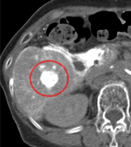
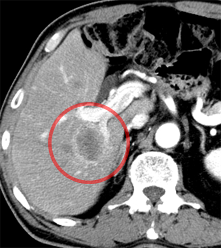
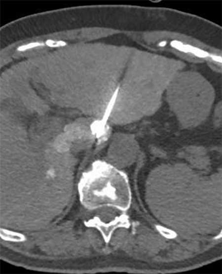
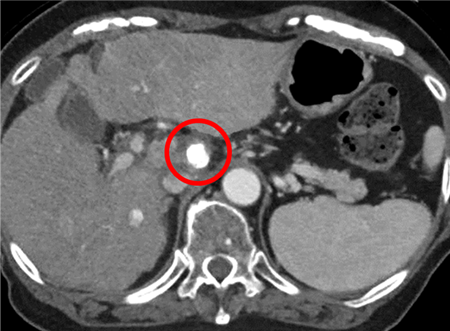
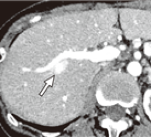
Liver cancer in a deep part of the caudate lobe, considered difficult to treat
Combined with catheter treatment
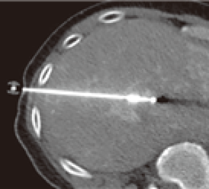
CT-fluoroscopy-guided RFA (Accurate puncture even in deep locations)
No recurrence for 5 years
As a result, depending on the patient's condition, we are able to flexibly consider a range of treatment options, including combination with surgery or chemotherapy.
Furthermore, for liver metastases from colorectal or rectal cancer, radiation therapies such as IMRT, proton beam therapy, and carbon-ion (heavy particle) therapy are also treatment options; however, depending on the tumor's characteristics and the condition of the lesion, sufficient effect may be difficult to achieve due to biological properties.
For this reason, it is important to select the appropriate treatment method for each case. At our clinic, we position IVR-centered local treatment as one effective option and actively pursue treatment aimed at achieving high local control.
The "Ablation Margin" That Determines Treatment Outcome
At our clinic,
we place great importance on ablating a somewhat wider, defined area around the tumor, in consideration of the possibility that invisible, latent cancer cells may exist not only in the tumor itself but also in the surrounding tissue—
this is what we call the "ablation margin."
This is expected to offer the following benefits.
A treatment strategy less dependent on tumor size or condition
Reduced risk of local recurrence
Improved local control
Evidence for the "Ablation Margin" (Reference)
Livraghi et al. Radiology 2000: Local control rate of RFA
Lencioni et al. Hepatology 2005: Indications and outcomes of RFA
Ahmed et al. Radiology 2011: Importance of the ablation margin
Gillams AR et al. Eur Radiol: Usefulness of CT-guided RFA
ð Many studies have shown that securing an adequate ablation margin plays a major role in reducing local recurrence.
Features of RFA at Our Clinic
In addition to the above, based on our extensive experience of more than 10,000 past cases, we perform the following treatments to further improve local control.
Addressing large tumors through combination with catheter treatment (embolization): By combining transarterial embolization (TAE), curative treatment can be expected even for large tumors exceeding 3 cm, which normally have limited indications. In addition, because treatment is performed while confirming the tumor's location and condition on the spot using catheter-based CT (such as CTHA or CTAP), it leads to more accurate and reliable treatment.
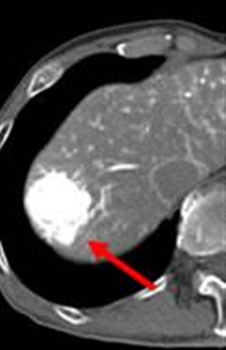
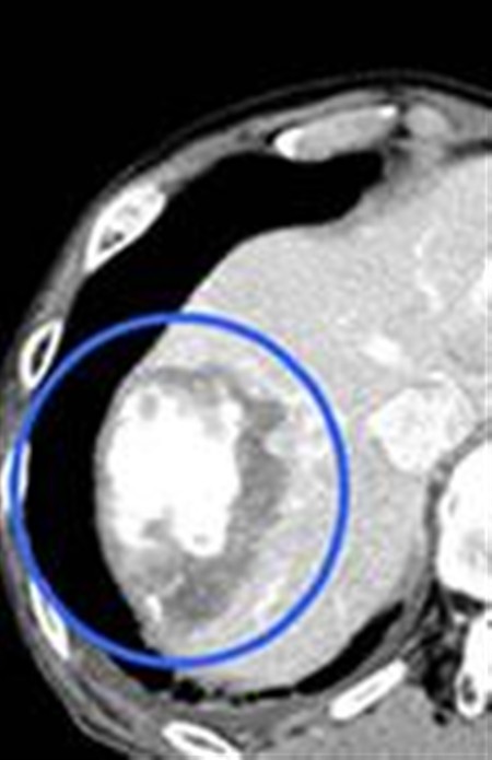
Before treatment (Large liver cancer)
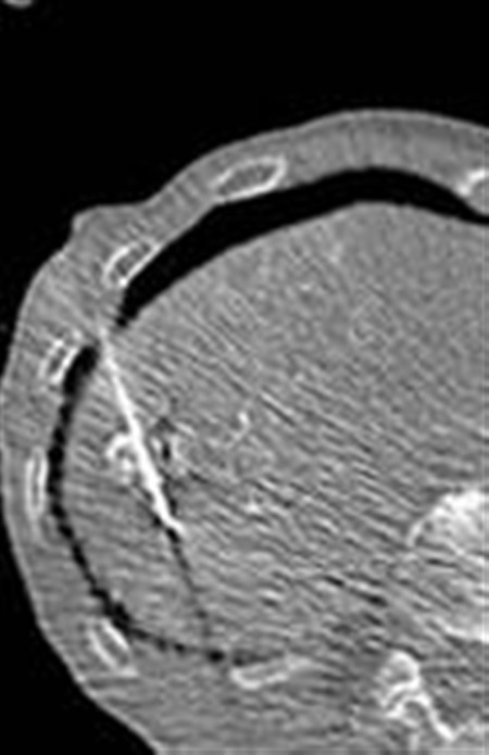
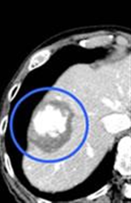
CT-fluoroscopy-guided RFA (Unaffected by gas bubbles)
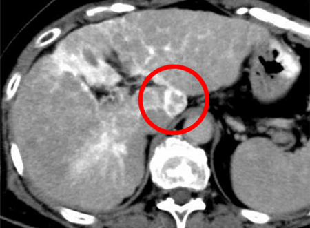
Immediately after treatment (healed) (The treated area appears outlined in black)
5 years after treatment (no recurrence) (The treated area appears outlined in black)
Before treatment CT-fluoroscopy-guided RFA Immediately after treatment (healed) 5 years after treatment (no recurrence) (Large liver cancer) (Unaffected by gas bubbles) (The treated area appears outlined in black)
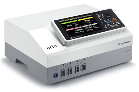
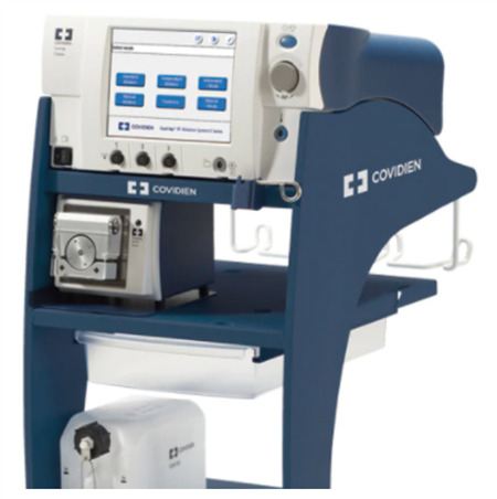
・Advanced systems and proven track record: At our clinic, we use multiple treatment devices as appropriate, providing treatment suited to each tumor's condition. In addition, procedures are performed by specialist physicians with extensive experience, delivering safer and more reliable treatment.
arfa
VIVA RF System
Cool-tip
In this way, using extensive expertise and a range of medical devices, we actively perform CT-fluoroscopy-guided radiofrequency treatment even for difficult-to-treat cases.
(References) Taku Yasumoto., et al. Innovative techniques for image-guided percutaneous puncture: Navigating complex cases for successful outcomes Interventional Radiology, 9(3): 99-111, 2024.
Treatment Eligibility
At our clinic, we do not categorically rule out treatment based solely on tumor size (over 3 cm), number (more than 3), location (near vessels, the gallbladder, or the gastrointestinal tract; beneath the diaphragm; or deep-seated), or reduced hepatic functional reserve. Even with many metastases, large tumors, or locations generally considered difficult, we judge eligibility by weighing how effective treatment is likely to be against the risks.
(For reference, the general standard indications are as follows.)
Primary liver cancer: Child-Pugh class A/B liver function, without portal vein invasion.
Metastatic liver cancer: Hepatic lesions 3 cm or smaller and no more than 3 in number.
Flexible Determination of Eligibility
Tailoring to each patient, we combine catheter-based treatment and RFA and also incorporate perfusion therapy, providing IVR for a wide range of lesions. We also treat liver metastases from other organs, such as colorectal cancer, and deliver care based on the latest knowledge in collaboration with university hospitals, research institutions, and nearby medical facilities.
We also provide appropriate treatment, based on accurate diagnostic imaging, for patients previously told that options were limited, that no effective treatment existed, that chemotherapy was not working, or who experienced repeated recurrence.
Transarterial Chemoembolization (TACE) for Liver Cancer
Transarterial chemoembolization (TACE) is a treatment in which a thin tube called a catheter is advanced to the blood vessels feeding the tumor; tumor growth is then suppressed by delivering anticancer drugs and temporarily blocking the blood supply. This technique—essentially starving the cancer of its blood supply—is one of the standard treatments now performed at many medical institutions.
At our clinic, we aim for more effective treatment by advancing the catheter as close to the tumor as possible. Precise techniques that selectively reach even the fine feeding vessels with a microcatheter have been scientifically shown to maximize treatment effect and contribute to longer survival.
TACE is also performed for liver cancer that has metastasized from other organs; however, because collateral pathways can develop after TACE alone, recurrence is sometimes seen. We therefore combine radiofrequency ablation (RFA) as needed to enhance the treatment effect.
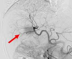
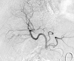
Before TACE (The dark shadow is the tumor)
After TACE (Shadow has disappeared, but there is a risk of recurrence)
Liver metastasis adjacent to the portal vein
CT-fluoroscopy-guided RFA after TACE
Complete ablation (CR) confirmed (No recurrence for 7 years)
We perform every procedure with careful attention to safety, but complications can occur in rare cases. Relatively common ones include temporary changes in liver function, fever, and pain. Most are transient and improve with oral medication or observation, and we respond appropriately according to each patient's symptoms. We will explain the details fully during your outpatient consultation.
Treatment of Liver Cysts
About Multiple Kidney and Liver Cysts
A fluid-filled, sac-like structure in the kidney or liver is called a "cyst." Cysts often increase with age; a single cyst is usually asymptomatic and simply monitored. When cysts are numerous (multiple cysts), however,
Organ enlargement
Abdominal bloating
Pressure symptoms
Pain
and other symptoms may appear. In polycystic kidney disease (PKD) in particular, reduced kidney function may also occur, requiring careful management.
General Treatment
Treatment of multiple kidney and liver cysts is determined by the presence and severity of symptoms.
Asymptomatic: Monitoring
Symptomatic:
Cyst puncture and drainage
Sclerotherapy (e.g., with ethanol)
Surgery (such as fenestration)
However, with multiple cysts,
Numerous cysts
Tendency to recur
Greater surgical invasiveness
are among the challenges.
Treatment for Multiple Liver Cysts at Our Clinic
At our clinic, for multiple liver cysts we perform image-guided percutaneous drainage (IVR).
This treatment involves
Confirming the position using imaging
Inserting a fine needle or catheter into the cyst
Draining the fluid
a minimally invasive treatment. We combine sclerotherapy as needed to help prevent recurrence.
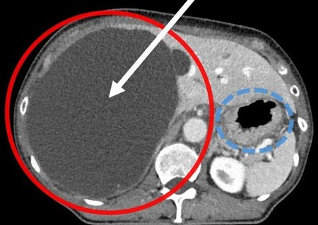
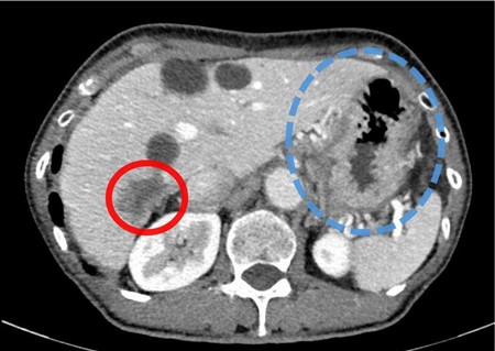
Giant liver cyst (red circle) Before treatment: the stomach (blue dotted line) is compressed
The liver cyst has markedly shrunk (red circle) 6 months after treatment: the stomach has expanded normally
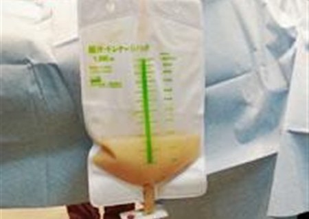
A thin tube is inserted to drain the accumulated fluid into a bag
Advantages of Drainage
Can be performed with only a skin puncture (minimally invasive)
Little burden on the body
Expected improvement of symptoms (such as abdominal bloating and pressure)
Same-day (outpatient) treatment possible
Short hospital stay even if admission is required
For patients with particularly strong cyst-related symptoms, this treatment is especially expected to improve quality of life (QOL).
Please Consult Us If You
Have abdominal bloating or pressure due to multiple liver cysts
Have cysts that are large and interfere with daily life
Wish to avoid surgery and prefer minimally invasive treatment
Have been treated elsewhere but your cysts have not shrunk
Were told to simply monitor the condition but are concerned about your symptoms
At our clinic, we combine IVR treatments such as CT-fluoroscopy-guided RFA and TACE to provide minimally invasive, precise treatment for liver tumors.
Even in cases where surgery is difficult, or cases involving recurrence or metastasis, we consider the possibility of treatment according to the patient's condition.
IVR Treatment for Portal Hypertension
What Is Portal Hypertension
When blood flow within the liver is impaired by conditions such as cirrhosis, the pressure in the portal vein (which carries blood from the intestines and spleen to the liver) rises. This is called "portal hypertension."
As portal pressure rises, collateral pathways (bypass vessels) that detour around the normal blood-flow route develop, causing various complications.
Representative examples include:
Gastric varices
Esophageal varices
Rectal varices
Ectopic varices such as duodenal varices
Hypersplenism
Thrombocytopenia
Pancytopenia
among others.
Treatment for Portal Hypertension
Treatment of portal hypertension aims to prevent bleeding, achieve hemostasis when bleeding occurs, maintain liver function, and improve cytopenias.
Endoscopic treatment or medication may be used, but depending on the condition, catheter-based IVR can be effective.
At our clinic, we perform various IVR treatments according to each patient's condition.
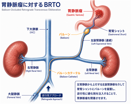
BRTO for Gastric Varices
What Are Gastric Varices
Gastric varices are varices that form around the stomach due to portal hypertension.
If they rupture, they can cause massive bleeding and may require emergency treatment.
BRTO is performed by inserting a catheter from a vein in the groin or neck and treating the varices while occluding the splenorenal shunt connected to them.
At our clinic, we perform BRTO for
Gastric varices
Splenorenal shunts associated with gastric varices
Large shunts causing hepatic encephalopathy
and other related conditions.
It is a minimally invasive treatment that can be done without open surgery, and we combine it appropriately with PTO and PSE (described below).
PTO for Ectopic Varices
What Are Ectopic Varices
Varices can also occur outside the esophagus and stomach.
Representative examples include:
Rectal varices
Duodenal varices
Small-bowel varices
Peristomal varices
among others.
Collectively, these are called "ectopic varices."
PTO (Percutaneous Transhepatic Obliteration)
PTO is a treatment in which a catheter is inserted into the portal vein via the liver to directly approach the bleeding varices.
At our clinic, we consider PTO according to the condition for
Rectal varices
Duodenal varices
Bleeding from ectopic varices
among other conditions.
PSE for Hypersplenism
What Is Hypersplenism
In portal hypertension, the spleen can enlarge and excessively destroy platelets, white blood cells, and red blood cells.
As a result, thrombocytopenia, leukopenia, anemia, and other conditions may occur.
PSE (Partial Splenic Embolization)
PSE embolizes part of the blood flow into the spleen to partially suppress splenic function.
It is expected to improve the platelet count and make it easier to continue chemotherapy and IVR treatment.
At our clinic, we perform PSE according to the condition for
Patients for whom continued treatment is difficult due to thrombocytopenia
Patients with pancytopenia due to hypersplenism
among other patients.
Please Consult Us If You
Have been diagnosed with cirrhosis
Have been told you have gastric varices
Have rectal or other ectopic varices
Experience repeated hepatic encephalopathy
Find it difficult to continue treatment because of a low platelet count
Have been told you have hypersplenism
At our clinic, we provide minimally invasive, IVR-centered treatment for the various conditions associated with portal hypertension. We consider a treatment plan tailored to each patient's condition, so please feel free to consult us.